Transmission EM
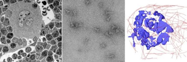
In transmission electron microscopy (TEM) an image is formed by a beam of electrons passing through specimens such as ultrathin sections of tissue/cells or material deposited on an EM grid. Contrast in the image arises from differential absorption of electrons by native or stained structures within the specimen.
The Wolfson Bioimaging Facility is exceptionally well equipped to support a range of TEM applications with its two high specification microscopes.
- The ThermoFisher Talos L120C TEM (new in June 2024) is a 120 kV TEM Suited for a range of sample types and applications from routine cellular or tissue level TEM through to electron tomography and cryo-TEM. Additionally, software available allows for single particle analysis (SPA), batch data collection and automated tile scanning.
- The FEI Tecnai 20 is a 200 kV TEM capable of high resolution imaging and can also be used to image thicker specimens, including creating tilt series for electron tomography. In addition it has cryo-TEM and scanning transmission electron microscopy (STEM) capabilities.
Further information is available on sample processing, technical specifications of the Talos L120C and Tecnai 20 microscopes and the GW4 facility for high-resolution electron cryo-microscopy.