Live cell imaging
Live cell imaging is a well-established speciality of the Wolfson Bioimaging Facility.
Each light microscopy mode offered by the Wolfson Biomaging Facility can be applied to live cells and almost all of our microscopes are equipped with incubators and CO2-enrichment facilities to enable environmental control.
Imaging techniques that require intense illumination inevitably affect some cellular functions and at worst can be highly phototoxic. For this reason widefield microscopy has advantages for many live cell experiments and especially for long-term imaging.
Specialised systems
In addition to traditional widefield microscopes we have two specialised automated live cell imaging systems that have advantages for long-term and high content live cell imaging.
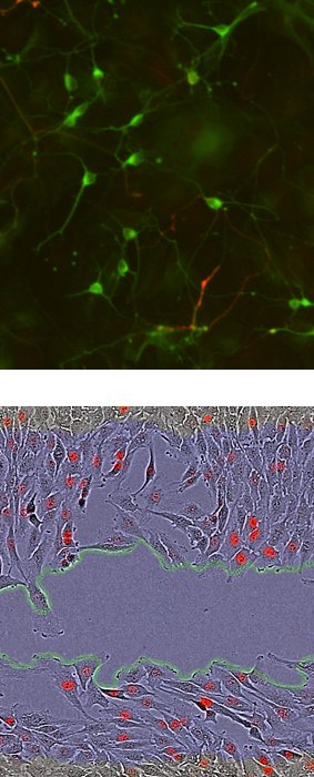
IncuCyte ZOOM
The IncuCyte ZOOM from Essen Biosciences is housed inside a CO2 incubator for optimised environmental stability.
The system offers the following key features:
- multi-sample imaging over hours and days
- range of magnifications – 4x, 10x, 20x lenses
- phase contrast imaging
- fluorescence imaging – red and green
- reproducible and repeated imaging of multiple fields - including whole well imaging
- imaging of up to six multi-well plates or combination of plates, flasks and dishes
- software tools for batch analysis ('high throughput' imaging) of cell number, confluence, cell fluorescence and morphology
- neuronal cell and dendrite analysis module
- hardware and software tools for scratch wound migration assays in 96-well format.
Incucyte Zoom technical specifications (PDF, 193kB)
Z Celldiscoverer 7
The Z Celldiscoverer 7 is an automated microscope incorporating environmental control.
The system offers the following key features:
- multi-sample imaging over hours and days
- wide range of magnifications – 2.5x-100x with automated lens switching and adjustment
- phase gradient contrast imaging
- fluorescence imaging – blue, green. Yellow, red, far red
- repeated imaging of multiple fields - including user-selected areas and whole well imaging
- imaging of one multi-well plate, 6 small dishes, 1 large dish or two slides
Celldiscoverer 7 technical specifications (PDF, 197kB)
Advice with Large file handling (PDF, 88kB)
Recent papers
- Edmunds., Wong, Ambler, Milodowski., Alamir, Cross, Galea, Wulfing, Morgan (2022). The adenosine 2A receptor and TIM3 directly inhibit the killing of tumor cells by cytotoxic T lymphocytes through interference with cytoskeletal polarization. Commun. Biol., 5, 9.
- Young, McGowan, Jepson, Adams (2020) Impairment of cell adhesion and migration by inhibition of protein disulphide isomerases in three breast cancer cell lines. Bioscience Reports 40: BSR20193271
- Bayliss, Sundararaman, Granet, Mellor (2020) Raftlin is recruited by neuroplin-1 to the activated VEGFR2 complex to control proangiogenic signaling. Angiogenesis 23: 371-383
- McNeill, Wray, Sala-Newby, Hindmarch, Smith. Ebrahimighaei, Newby, Bond (2022) Nuclear actin regulates cell proliferation and migration via inhibition of SRF and TEAD. Biochim Biophys Acta Mole Med Cell Res 1867:118691
More information and access
For further information or to arrange access to this equipment, contact one of the team.
We welcome comments or suggestions. Please contact one of the team or one of the advisory group.