Lightsheet
Lightsheet fluorescence microscopy (also known as single plane illumination microscopy; SPIM) enables 3D imaging of relatively large specimens by sequential illumination of single planes.
Reduced light exposure compared to conventional imaging modes limits photodamage and has made lightsheet microscopy a preferred option for in vivo imaging of model organisms such as Zebrafish and Drosophilla. Deep imaging can be further enhanced in lightsheet microscopy by acquisition of images at multiple angles before recombining to form a 3D dataset.
The Wolfson Bioimaging Facility’s Zeiss Z.1 lightsheet system arrived in February 2018 following the award of a Wellcome Trust multi-user equipment grant. This is a multi-laser (405, 445, 488, 515, 561 and 638nm), turn-key system capable of imaging a broad range of samples, which are typically embedded in 1% low-melting agarose within glass capillaries. The Wolfson Bioimaging Facility support team can assist with sample preparation, image acquisition and image processing.
Lightsheet image collection is via two high resolution sCMOS cameras. Flexible acquisition options are provided with a range of filter modules and objective lenses (5x, 10x, 20x and 40x). The lightsheet system is equipped with environmental control via temperature and CO2 incubation. An additional 20x lens and specialised chamber enable imaging of cleared tissue (RI ~1.45). In addition to the acquisition workstation, which provides automated imaging through Zeiss Zen software, a second analysis workstation provides advanced 3D/4D rendering with Arivis Vision4D software.
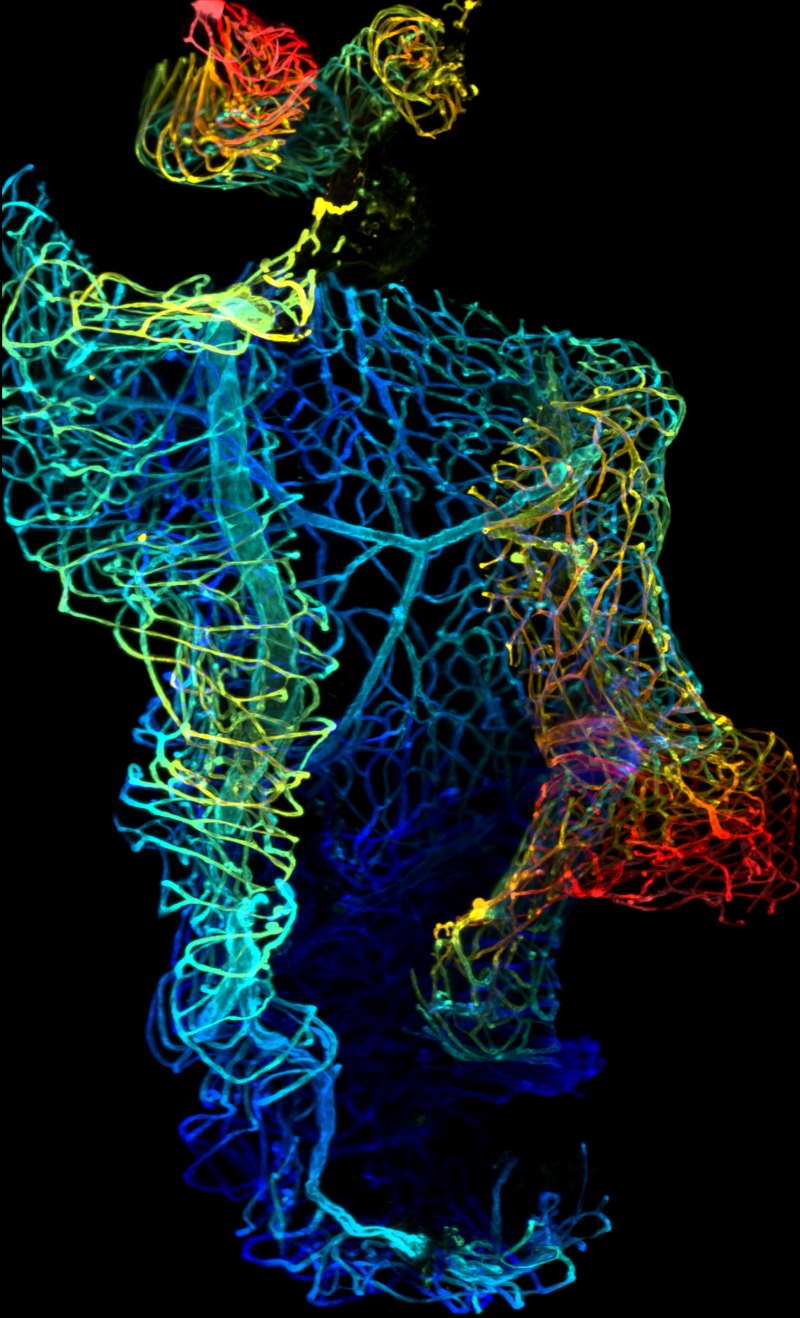
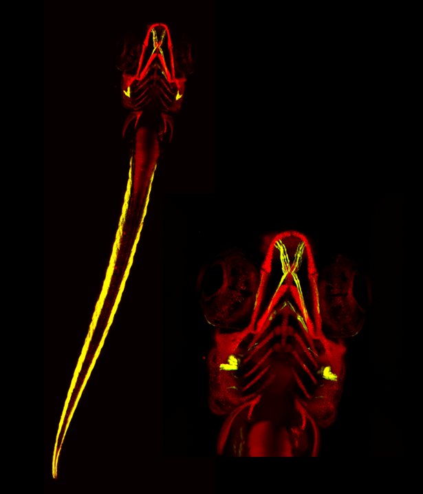
Human cerebral vasculature in fixed and cleared post-mortem tissue. Acquired by Katy Jepson (Wolfson Bioimaging Facility, University of Bristol) and Robbie Fisher (Translational Health Sciences, Bristol Medical School).
Lightsheet technical specifications (PDF, 94kB)
Advice with Large file handling (PDF, 88kB)
First paper including lightsheet data:
- Tuffin, Chesor, Kuzmuk, Johnson, Satchell, Welsh, Saleem (2021). GlomSpheres as a 3D co-culture spheroid model of the kidney glomerulus for rapid drug-screening. Commun. Biol. 4(1):1351.
More information and access
For further information or to arrange access to this equipment, contact one of the team.
We welcome comments or suggestions. Please contact one of the team or one of the advisory group.