Bioimaging at the cellular level
The beauty of imaging cells and cellular structures is combined with powerful image analysis methods in the University’s Wolfson Bioimaging Facility. Paul Verkade, Head of the Electron Microscopy (EM) unit in this Facility has combined his own expertise on cellular trafficking, with image processing research in BVI (Alin Achim, David Bull, Paul Hill and Pui Anantrasirichai) to further our understanding of cell structures and dynamics.
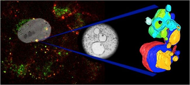
The work of Verkade’s group is world-renowned for its work on Correlative Light Electron Microscopy (CLEM), where the history of an event in live light microscopy is combined with high-resolution structural EM data of that same event. Mapping those two datasets onto each other has been a major challenge.
Automated registration and mapping software developed within BVI has recently transformed the way we can perform CLEM experiments. Other example contributions include analysis of the 3D ultrastructure of insulin granules inside islets of Langerhans and the identification of fusion and segregation events inside cells by live light microscopy. As our microscopes continue to produce ever more information-rich data, the analysis and understanding of this data will rely even more heavily on advanced image processing methods.