|
-
Peripheral Nuclei
-
Massive Cytoplasm
 |
Figure 1 - Skeletal muscle Longitudinal section. Bar is 250 microns |
At low magnificaiton, skeletal muscle in longitudinal section is non-branching and can be identified by peripheral nuclei. The large white vertical lines are knife marks from sectioning (artifact). You will not be able see striations clearly with less than 40x objective and only clearly with 100x oil-immersion lens.
===========================================================
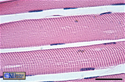 |
Figure 2 - Skeletal muscle longitudinal section at high magnification. Bar is 30 microns |
At higher magnification, the characteristic striations become visible. I-bands (isotropic) are light while A-bands (anisotropic) are dark. Notice the very obvious dark inclusions at the edge of the fiber! These are the nuclei - rather pushed to the periphery of the cell, hence peripheral nuclei. This is a very distinctive feature of skeletal muscle.
===========================================================
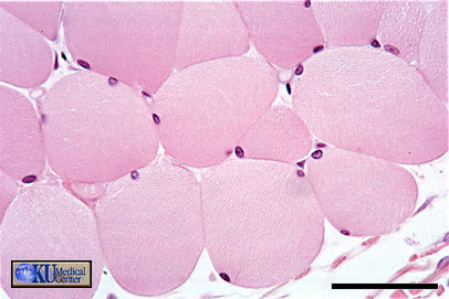 |
Figure 3 - Skeletal Muscle transverse section. Bar is 50 microns |
In cross section, skeletal muscle is identified by peripheral nuclei and large amounts of cytoplasm (or small extracelluar space!). Occasionally this feature can be obscured if the fibres have shrunk excessively during fixation and processing.
===========================================================
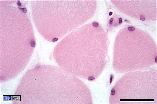 |
Figure 4 - Skeletal muscle transverse section (Bright field illumination). Bar is 30 microns |
Compare this slide to the one below (slide 5). They are photomicrographs of the same section but the optical method differs. Figure 4 (above) is taken with normal bright-field illumination (the same sort of illumination that you will use)
===========================================================
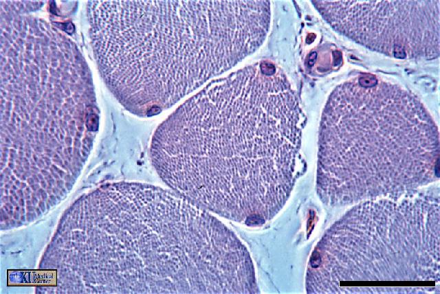 |
Figure 5 - Skeletal muscle transverse section (phase-contrast optics). Bar is 30 microns |
Same slide as the slide above (4), but this time viewed using phase contrast optics. Notice how the individual myofibrils that make up muscle fibers show up more clearly (look particularly at the fiber on the left of the image and you should see a sort of mosiac pattern).
===========================================================
|