Holographic microscopy
Digital Holographic Microscopy (DHM) is a new quantitative method for phase contrast microscopy. This is a technique that measures the differences in the distance that light travels through objects, and can do so with the a resolution of 1 nanometer, allowing you to measure quantifiable 4D tomographic changes in your sample. The method is truly non invasive, it is non contact (unlike electrophysiology) and there is no need to use fluorescent dyes. Digital holographic microscopy can be used as an optical patch clamp, for example, to quantifiably measure channel activation, via ion / water flux measurements.
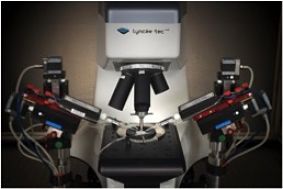
BrisSynBio Digital Holographic Microscopy systems
Multi-wavelength platform
A 4-wavelength laser source (445, 488, 515 and 640nm), 10x, 20x, 40x and 100x oil immersion Leica fluotar objectives, a polarization module and a 25 fps multiwavelength DHM camera.
Dual-wavelength platform
A 2-wavelength laser source (666 and 794nm), 5x, 10x, 20x, 40x, 63x and 100x oil immersion Leica fluotar objectives, an Epi fluorescence system, and a 1000 fps dual-wavelength DHM camera (100 fps if fluorescence is used).
The DHM suite is located in the Life Sciences Building and is charged using a "consumables-only" model.
Academic Equipment Lead: Dr Nicholas Roberts
Equipment booking
BrisSynBio users are welcome to book our equipment.
Please contact us (brissynbio-equipment@bristol.ac.uk) for more information about this equipment.