Contents | 1 | 2 | Previous | Next

1. EC Coupling 1. At the NMJ Logo


There are not too many problems and the general sequence of events appear to be reproduced well enough in tutorial and examination, but some students mix up some of the channels and receptors, perhaps placing things that belong on the post-juctional membrane on the pre-junctional membrane. This type of thing is a problem to an examiner because they have to decide whether it is just an error or if it represents a more serious lack of understanding. So, the problems thrown up are partly to do with the sequence of events at the pre-junctional and post-junctional membranes and partly to do with the specific structures, receptors and channels that play a part in the whole event.


A cartoon of a neuromuscular junction (seen as illustration in lecture 5.)

Figure 1 - A cartoon of a neuromuscular junction (seen as illustration in lecture 5.)


The pre synaptic membrane releases vesicles of transmitter from special areas called ‘dense bodies’ or active zones. These are aligned with infoldings or ‘clefts’ in the post-junctional membrane.

  • This may be to maximise the efficiency of the junction so that fewer vesicles are needed.
  • In Man (many deep clefts) only ~20 vesicles are released per AP.
  • The top of the clefts are packed with nACh receptors, the bottom half has only voltage gated Na+ channels.


Cartoon of section of NMJ  to illustrate the alignment of dense bars (site of transmitter release) and the tops of the junctional clefts (site of greatest nAChR density)

Figure 2 - Cartoon of section of NMJ to illustrate the alignment of dense bars (site of transmitter release) and the tops of the junctional clefts (site of greatest nAChR density)


From nerve action potential to muscle action potential Top


The sequence of events is relatively simple but you do need to pay attention to the particular channels and receptors that play a role in the transmission of excitation from nerve to muscle.
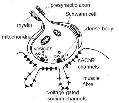
  • An action potential, having propagated along the axon of the alpha motor nerve, from the ventral horn of the spinal chord to the muscle, invades the pre-junction membrane or end plate.
  • The depolarisation (voltage changing from negative towards zero and even becoming positive to zero) caused by the invading action potential is detected by voltage-gated calcium channels which open, admitting calcium ions, raising the concentration of Ca2+ within the end plate and causing release (calcium-dependent mechanism) of about 20 transmitter vesicles.
  • The transmitter diffuses from the pre-junction release site to the post-juctional membrane, a distance of some 20 nm.
  • ACh binds to nicotinic acetylcholine receptors (nAChR) (2 molecules of ACh per receptor), causing it to open. Each action potential in the motor nerve releases sufficient ACh to open about 50, 000 receptor channels, about 0.5% of the available receptors.
  • The channels can pass both K+ and Na+ ions, but in reality the ions of only one species moves in any quantity.
  • The movement of ions tends to push the membrane potential of the post-junctional membrane towards 0 mV. This is detected by voltage-gated Na+ channels in the bottom of the clefts. The Na+ channels open which increases the amount of depolarising current (current density) flowing in the junctional area.
  • The current flowing in the area of the NMJ is sufficient to cause local current flow and depolarisation of the surrounding membrane, activating the voltage-gated Na+ channels and initiating an action potential which then propagates away from the NMJ to depolarise the entire muscle fibre.


Contents | 1 | 2 | Previous | Next
Copyright © 1998 University of Bristol. All rights reserved.
Author: Phil Langton
Last modified: 9 Jun 1999 20:12
Authored in CALnet