Learning anatomy online is to benefit from a new tool using the latest technology, which allows users to see real specimens in high-definition 3D.
The free online resource, 'real 3d anatomy’, will offer students high-definition 3D anatomical models to help supplement their study when away from the lab.
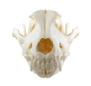
Using images of real specimens, the 3D models comprising approximately 20 individual bones from a canine skeleton are fully rotatable in 360 degrees from any angle and are complete with zoom functions.
Created by a team from the University of Bristol, led by Dr Cathy Fuller and Adam Baumberg from Creative Dimension Software Ltd (CDSL), the interactive resource has been produced using photographs of a canine bone set which are then transformed into 3D models using specialist software.
Dr Fuller from the University’s Centre for Comparative and Clinical Anatomy (CCCA), said: “The tool is aimed at anyone, anywhere, who is studying or has an interest in anatomy and who may have no access to real bones. We hope that the interactive 3D nature of this resource will provide people with the next best thing.
“What’s special about this resource is that although other online 3D anatomy resources are available, most use computer-generated images. The specialist software we have used in creating the resource means that the anatomical models are created from images of real specimens, and in addition are freely rotatable as if the user is holding the specimen in his or her own hand.”
The project has been funded by the University’s Centre for Comparative and Clinical Anatomy, which delivers undergraduate teaching in anatomy to preclinical medical, dental and veterinary students as well as to undergraduate BSc students from a variety of disciplines in the medical sciences and elsewhere in the University.
Dr Fuller added: “We hope to be able to provide more models like this, however we are now at the point of needing funding in order to develop this further.”
The free resource is available on the ‘real 3d anatomy’ website. Users with a standard browser (Flash enabled) can view an interactive 3D presentation of the complete canine skeleton as well as a demonstration showing the thoracic organs of the dog (the chest cavity with heart, lungs and ribs).