
By using biophysical, biochemical and molecular biological techniques, it is possible to study how changes in the mechanisms that regulate these ion channels can lead to the development of arrhythmias and other types of heart disease. For example, in heart failure, contraction of the heart is much weaker than normal, causing blood to be pumped less effectively. This is due partly to less calcium entry into the cell because of changes in the function of ion channels, which may be at least partly due to the cells having fewer t-tubules.
Synchronised contraction of millions of cells causes the heart to beat
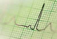
Activity of ion channel behaviour that may lead to arrhythmias can be detected by an electrocardiogram (ECG). Increased risk of one particular type of arrhythmia is caused by impairment of a potassium channel – the ‘HERG’ channel – which causes a change picked up by the ECG known as ‘long QT syndrome’. Surprisingly, a number of drugs in routine use for non-heart related problems were found to cause this abnormality, so these were subsequently withdrawn. The drugs acted by blocking the HERG channel. A key question that researchers at the Bristol Heart Institute are now trying to answer is how the structure and function of this particular ion channel makes it susceptible to being blocked by such drugs. Answers to this problem are clearly important for the future design of safe drugs.
Major differences in men's and women's cardiac arrythmias
Interestingly, there are major differences between men and women in the likelihood of their suffering serious cardiac arrhythmias – women are more susceptible to arrhythmias due to long QT syndrome, whereas men appear to be more susceptible to arrhythmias arising from abnormal function of other ion channels. But astonishingly little is known about the mechanisms that cause these gender differences. It seems probable that they arise from the actions of the different sex hormones – testosterone and oestrogen – on the heart. Investigating the role of the sex hormones in modifying cardiac ion channel behaviour should uncover the basis of these sex differences in the susceptibility of the heart to arrhythmias. In turn, this will allow the development of drug therapies that are equally safe for both men and women.