
According to the news, we binge drink, smoke too much, eat the wrong foods and don’t take part in physical activities as often as we should do. We’re overweight, diabetic and our blood pressure and cholesterol levels are through the roof. It’s no wonder then that cardiovascular disease (CVD) is the main cause of death in the UK.
About half of the deaths from CVD are from coronary heart disease (CHD), which incorporates conditions such as angina and heart attacks. Whereas morbidity from CHD appears to be on the rise within the UK, death rates have been falling steadily since the 1970’s.
So why have the death rates been falling?
As with any disease, if you are aware of its physiological effects and how and when it presents itself, then it becomes more easy to detect and diagnose, thus giving more time to treat. So if a patient presents with chest pains, how can one further investigate this to make a sound diagnosis?
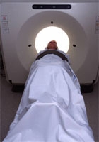
One approach is to use medical imaging techniques, such as angiography and computed tomography (or CT/CAT scans). These work by injecting a contrast medium into the patient, which essentially gives tissues and surrounding fluids (such as blood) different opacities.
When exposed to X-ray radiation, the differing opacity between the tissues and fluids means they will absorb the X-rays at different rates. Computerised detection systems or X-ray film are then used to detect the varying intensities of X-rays that pass through the tissues and an image of the body part, for example the heart, can then be produced.
At present, invasive coronary angiography is the gold standard investigation to detect problems associated with CHD, such as narrowing of the arteries. This involves puncturing an artery and inserting a catheter through the aorta towards the coronary artery so that the contrast material can be injected.
Due to its invasive nature however it carries a small risk of serious complications. New technologies such as multi-slice computed tomography (MSCT) use special X-ray equipment and an array of detectors to measure the X-ray profiles that are then computer-processed to give a cross section of tissues and organs and even produce a 3D image. MSCT scans still require an injection of contrast material but are much less invasive.
Why not use CT scanning as investigative approaches all the time?
Due to the tremendous amount of data acquired and processed in CT scanning, in the past it often took as long as 12 hours to produce a final image. Advancements in computed tomography over the last ten years however, have allowed 3D images of tissues and organs to be produced in real-time.
A major concern of this procedure, one that is being addressed by Dr Andreas Baumbach in the Cardiology department of the BRI, is the dose of radiation that is required to produce such an image.
As a general rule for CT scans, the higher the radiation dose, the higher the resolution and the better quality the final image. Even though technological advancements have improved the efficiency of the scanners, radiation doses are still classed as moderate to high and there is still pressure, especially in the U.S, to increase the radiation dose for higher resolution imaging.
Higher radiation doses are of course dangerous and can increase the risk of inducing a fatal cancer. What would then seem to be a necessary study, but until now had not been comprehensively assessed, is the effective radiation dose that a patient receives when having a CT scan.
Baumbach and colleagues, working closely with the Department of Physics, have quantified and compared effective doses from conventional angiography and MSCT coronary angiography.
In a patient population of 180, all with suspected coronary artery disease, estimates of the effective dose were derived from expose data recorded for each patient examination. After running the data though some statistical calculations the resultant figures showed that the effective dose of X-ray radiation received when undergoing MSCT was significantly higher than that for conventional angiography.
With considerably higher radiation doses and a higher chance of inducing a fatal cancer, when should CT cardiac imaging be used over conventional angiography?
Although conventional angiography carries invasive mortality and morbidity risks, fifty percent of the procedures are performed on patients with acute problems, in which case the benefits outweigh the risks. In these situations MSCT scans are not as effective in aiding with a diagnosis as the standard angiograms; what they are very good at doing however, is displaying normal conditions.
The CT angiography is an excellent tool to exclude problems in the coronary arteries in patients with symptoms, but not a very high likely hood of coronary blockages. A further emerging indication is the scanning for coronary calcification, which is an early marker for CVD. This can add to the risk profile of the asymptomatic patient and can trigger the need for further investigations or the start of preventative treatment.
This risk-to-benefit ratio is one that Dr Baumbach and colleagues are helping to define by assessing and comparing radiation doses between such cardiac imaging technologies. With MSCT scanners becoming increasing available, in conclusion to his research, Baumbach suggests that operators of the scanners must be aware of the radiation dose and the factors that affect it.
And for CT scanning and similar emerging cardiac imaging technologies fully to replace invasive procedures, then not only does the technology need to move on, but it needs to do so producing higher quality images using less radiation.
Dr Andreas Baumbach/Clinical Science at South Bristol