EM Labs
Contact: Dr Ben Buse and Dr Stuart Kearns
Electron Microbeam

Scanning electron image of chemical zoning within a plagioclase crystal from Mt St Helens. Zoning is important for understanding volcanic eruptions and diffusion studies, see Kate Saunders study
The electron microbeam laboratories provide a service to the Earth and Environmental science community as well as to all departments within the University of Bristol. The laboratories consist of two scanning electron microscopes SEM) and two electron probe microanalysers (EPMA), which enables us to provide a full range of electron and x-ray imaging, together with in-situ chemical analysis of both major and minor elements heavier than carbon.
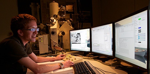
Scanning Electron Microscopy
The laboratories operate two scanning electron microscopes (SEM), one aimed at undergraduate projects and teaching and one aimed at other research projects. The laboratories have the Alicona MEX software for stereographic measurement.
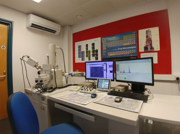
Thermo Scientific Apreo 2 SE

The laboratories operate a Thermo Scientific Apreo 2 SEM, installed January 2023, providing high performance for research. The microscope is fitted with a field-emission source providing 1 nm resolution. The instrument is equipped with multiple detectors, including a rapid inlens backscatter detector allowing fast sample imaging or simultaneous collection with cathodoluminescence signal. It also includes a conventional backscatter detector for highest definition. The instrument has a range of charge mitigation strategies permitting high resolution analysis of uncoated non conductive materials at high vaccum and were necessary has a low vaccum mode. The instrument is equipped with two Oxford Instrument energy dispersive detectors (EDS) providing rapid x-ray mapping and non-normalised standardless quantification. Samples up to 110 mm can be accommodated, with stage movement in x, y and z, rotation and 105° tilt.
Hitachi S-3500N SEM
The school operates a Hitachi S-3500N aimed at undergraduate teaching. It is a variable pressure microscope equipped with Thermo Noran energy dispersive spectrometer (EDS x-ray detector). This allows both coated (gold or carbon) or uncoated specimens to be quickly and routinely imaged and analysed.
The microscope is fitted with a tungsten filament which produces a stable and high beam current, making it ideal for x-ray analysis. Ultimate resolution is better than 10 nm at 25 kV, using the secondary electron (SE) detector. The instrument is equipped with a backscatter electron (BSE) detector which allows imaging of specimens that have a mean atomic mass change of ~>0.1.
The machine may also be used in low vacuum mode (>200Pa) in conjunction with the environmental secondary electron detector (ESED) and BSE detector. This enables electron imaging of both uncoated and hydrated samples. The machine is also equipped with a Peltier cold stage, which can be used to cool samples in situ to –30oC.
Irregular shaped samples may be accommodated, with the stage allowing movement in the x, y and z directions, as well as rotation and up to 60o of tilt at moderate working distances. The EDS detector (ThermoFisher Scientific) provides both standardless quantitative analysis and advanced x-ray mapping capabilities of elements heavier (and including) carbon.
Detection limits are in the order of 0.5 wt% (depending upon element and operating conditions) and analytical volumes around 2 to 3 cubic microns (again, dependant upon operating conditions).
All images are saved as high resolution bitmaps . Samples may be uncoated, carbon or gold coated. Facilities to carbon or gold coat are housed within the facility.
Electron Probe Microanalyser
The Electron Probe Microanalyser (EPMA) is a non-destructive technique for the analysis of polished and carbon-coated specimens, with some exceptions: conductive metals, and silver-coated carbonates. Analyses can be made at individual points (usually 1 to 40 microns in diameter) or over small (usually < 1mm2) areas of the sample surface to produce geochemical maps. Using the instruments at Bristol, almost every element of the periodic table can be determined with the exception of those with atomic numbers lower than boron.
Specimens are introduced into the sample vacuum chamber and viewed under high magnification to select areas of interest. A beam of electrons is fired at the sample surface producing X-rays with energies and wavelengths specific to the elements present. Instrument calibration is carried out by analysing standards with known compositions. An example of the use of EPMA is in the study of subtle chemical zoning in minerals by element mapping, which is often invisible using light microscopy.
The School has two EPMAs a JEOL JXA8530F Hyperprobe and a CAMECA SX100.
JEOL JXA8530F EPMA
The JEOL JXA8530F EPMA is equipped with a Field Emission Gun electron source and thus provides the highest possible spatial resolution. The instrument has 5 wavelength dispersive spectrometers (WDS) and a energy dispersive spectrometer (EDS) and is equipped with both the JEOL and the Probe for EPMA software https://probesoftware.com/, providing additional features such as mean atomic number backgrounds and time-dependent intensities. The JEOL software permits mapping of irregular areas. The instrument is equipped with a Peltier-cooled cold finger to assist in reducing contamination when analysing at low voltage or low overvoltage to achieve high analytical resolution. The samples can either be 1-inch rounds or thin sections provided then don’t have cover slips.
CAMECA SX100

The Cameca SX100 microprobe is equipped with 5 wavelength dispersive spectrometers and a tungsten source, providing high precision and accuracy at high accelerating voltages (15-20kV), corresponding to several microns depending on density.