Glomerular physiology
Glomerular water permeability assay
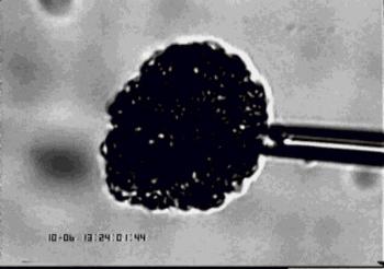
This allows us to measure the water permeability (LpA) of a single isolated glomerulus (from animals and humans) by fixing the glomerulus in place using a suction micropipette and changing the osmotic concentration of the perfusate. This can be done on glomeruli incubated in different stimuli for acute effects or glomeruli isolated from different animal models/human pathologies. This is an extensive refinement of a very powerful, yet not widely used, method for measurement of glomerular ultrafiltration coefficient.
Figure 1. Isolated Glomerular ultrafiltration coefficient (LpA) assay. Following isolation, glomeruli are incubated at 37°C in 1% BSA solution (with or without test solutions). Glomeruli are then individually loaded onto a suction micropipette. Flowing perifusate containing 1% bovine serum albumin (BSA) is rapidly switched to an oncopressive perifusate containing 8% BSA, eliciting transglomerular fluid efflux. The rate of the resultant reduction in glomerular volume is used to calculate glomerular LpA (Salmon et al, 2006).

Glomerular albumin permeability assay
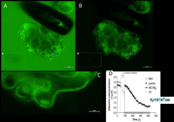
This assay is unique to our group and has recently been characterised and validated. As an extension to the assay above we can now measure relative albumin permeability across a single capillary loop of an isolated glomerulus. This is a fluorescence assay which involves pre-perfusing the kidney with a red cell membrane label (R18, to define capillary loops) and green labelled albumin (AF488-BSA). Using confocal microscopy we can measure the decline in fluorescent intensity in a capillary loop over time and from this and other parameters infer relative solute albumin permeability (Psalb).
Fig 2. Glomerular albumin permeability assay. 
A single rat glomerulus was perfused with labelled-albumin, and incubated in a bath containing labelled albumin (A). The bath was then rapidly switched to unlabelled albumin (B) and then the diffusive albumin solute permeability (PSalb) was calculated from the fluorescence decay in a single capillary loop (C) compared to a region in the bath (green square). PSalb was 7.9*10-7cm/s (D) (Desideri, Foster & Salmon, unpublished).
