SBF - SEM
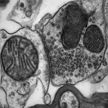
Serial block face scanning electron microscopy (SBF-SEM) enables high resolution 3D imaging of biological samples that have been fixed, stained and embedded in resin. It is also applicable to other materials that can be sectioned using an ultramicrotome. SBF-SEM can provide nanometer level ultrastructural information over relatively large fields of view – up to several microns in X,Y and Z.
SBF-SEM typically relies on an ultramicrotome being installed inside an SEM. The SEM collects sequential images of the block face while the ultramicrotome incrementally removes surface layers. The resulting images can be segmented and reconstructed to provide 3D data.
A BBSRC ALERT17 grant funded SBF-SEM (Zeiss GeminiSEM 450/Gatan 3View) was installed in the Wolfson Bioimaging Facility in February 2019.
Recent publications including SBF-SEM data:
Irwin, Williams, Speiser, Roberts (2022) The marine gastropod Conomurex luhuanus (Strombidae) has high-resolution spatial vision and eyes with complex retinas. Journal of Experimental Biology 225(16):jeb243927.
Laundon, Chrismas, Bird, Thomas, Mock, Cunliffe (2022) A cellular and molecular atlas reveals the basis of chytrid development. Elife 2022 Mar 1;11:e73933
More information and access
For further information or to arrange access to this equipment, contact one of the team.
Please fill in this form to register sample processing requests.
We welcome comments or suggestions. Please contact one of the team or one of the steering group.