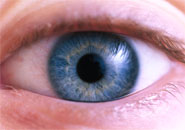
Using the new technique called ‘TEFI’ (Topical Endoscopic Fundal Imaging), Professor Andrew Dick, David Copland and the team from the University of Bristol’s Academic Unit of Ophthalmology, monitored changes in mice retina over time, without distress to the animals or the need for anesthesia.
The study focused on a condition in mice similar to human posterior uveitis, an inflammation that affects the back of the eye and which can be difficult to monitor using existing techniques. TEFI allowed the researchers to see changes to the eye that were previously undetectable.
“TEFI enhances our monitoring of clinical disease in a rapid and non-invasive fashion,” Copland said. “It will aid in the design of experimental protocols according to clinical observations.”
Professor Dick added: “Combined TEFI and histological methods enable the observation of clinical features and severity of disease, but information regarding the dynamics, phenotype, function and quantity of cellular traffic through the eye is only provided through detailed analysis of cell populations present in the eye at various stages of disease progression.”
The study, “The Clinical Time-Course of Experimental Autoimmune Uveoretinitis Using Topical Endoscopic Fundal Imaging with Histologic and Cellular Infiltrate Correlation,” was published this week in Investigative Ophthalmology and Visual Science. It featured the use of Topical Endoscopic Fundal Imaging (TEFI), a technique that uses an endoscope with parallel illumination and observation channels connected to a digital camera.