X-ray tomography (XTM) Facility
The Palaeobiology Research Group hosts the Faculty of Science micro-CT X-ray tomography facilities in the Life Sciences Building. This facility is available for all users in the Palaeo group, as well as to both other members of the University and to external users.
X-ray tomography facilities
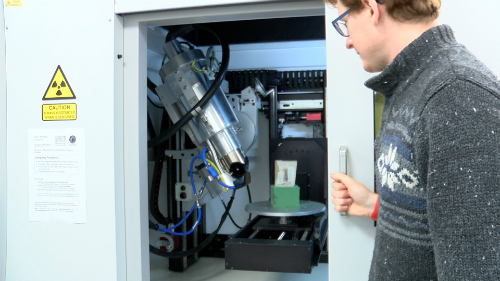
We have a Nikon XTH 225ST X-ray tomography scanner. This Computed Tomography (CT) system is suitable for a wide range of materials and sample sizes. It is used widely at Bristol for visualisation of fossil specimens in rock matrix, for biological specimens, or for inspection of geological rock samples, amongst other applications. View specifications and further information.
The scanner has three interchangeable x-ray sources suited for different purposes that are rotated through an approximately fortnightly cycle depending on demand:
- Reflection target: 225kV general purpose target suitable for range of samples and materials with minimum spot size of 3um.
- Rotating target: 225kV target that allows greater power for given spot size, suitable for large denser material, as well as for small specimens that are relatively dense (e.g. fossil/rock material). Minimum spot size = 10um.
- Transmission target: 180kV target suitable for the smallest samples (approx. 1–10mm) allowing spot size down to 1um.
Software
The scanner uses Nikon’s own software for scan acquisition and reconstruction of tomographs from the projections.
Additionally we have a single licence for VG Studio MAX that we use to visualise datasets, reorientate slices, and export datasets in a variety of widely compatible image datasets.
In association, we have an Imaging Lab on level 1 LSB with a range of high-powered workstations capable of running big datasets, and with network license allowing simultaneous users on Avizo and Dragonfly softwares for visualisation and segmentation of 3D models.
Output
Typical output from the scanner is .tif stacks of image files (cross-sectional slices revealing internal structures of the specimen), though other outputs are available upon request. Only raw imaging data is provided. Further analysis is the responsibility of the researcher/customer.
Resolution
The resolution of scans depends primarily on the distance from the object to the source of the x-rays.
For larger specimens (~20cm+ across) resolution will be in the region of 60-100microns, while for the smallest specimens (1-2cm or less) resolution is possible down to 1-3micron voxel size.
Specimen Dimensions
Maximum object dimensions (for single scan) are approximately 30cm in each of xyz dimensions, although the chamber is large enough to hold larger objects which may be scanned in multiple stacks (up to 50cm in diameter and 100cm in height).
Costs
The XTM Facility is a seperate Facility within Earth Sciences.
Costs for internal users are currently set by finance at £46.99/hr. To use our Imaging Lab, there is also an associated cost of £8.91/day. Please contact us for more details.
For external users please email for an official quote - for academic users costs are typically £60/hr + VAT and may incur extra operating time if scans must be done by one of our authorised users. Industrial users should email for more information.
Safety
The system is manufactured to IRR99 and is lead-shielded to <0.5uSv/hr with multiple interlocks to prevent accidental exposure to radiation.
There would be very high doses of x-ray radiation within the cabinet itself, but in the room the levels of radiation are below that which would require personal monitoring for exposure.
The room is a ‘supervised area’ with radiation levels monitored twice a year.
The radiation protection supervisor for x-rays is Liz Martin-Silverstone. Full documentation on safety protocols, including local rules, is available on request.
Facility Acknowledgement
All users agree to acknowledge the XTM Facility in any publications arising from data collected at the Facility. This includes (but is not limited to): a) mentioning the Facility by name (we suggest “CT data were obtained at the XTM Facility, Palaeobiology Research Group, University of Bristol”) in the methods when describing the scans; b) acknowledging the Facility and any technical support that assisted in data collection, if applicable within the acknowledgment section; and c) considering acknowledging technical support as a named author if the technician has significantly contributed to method development, analysis, or support, to be discussed through the duration of the project.
Facility users are entitled to one (1) day of technical support either through training or having the scans done for them. Any additional time needed must be compensated by either paying for additional time on the scanner or through authorship on any publications using scans performed by the Facility Manager.
Quotes, booking and information
For further information or enquiries, email the Palaeobiology Lab Manager Dr Liz Martin-Silverstone at palaeo-lab@bristol.ac.uk, or alternatively fill out this booking form and we will get back to you as soon as we can.
If you are requesting a quote, provide the following information:
- what the specimen is (type(s) of material)
- number of objects to be scanned
- approximate size (h x w x d)
- what you are interested in seeing in the sample (approx. size of known features etc.)
- approx resolution required (if known)